
Pollen
Roll the mouse over a picture to see the full resolution image.
Acknowledgment: Louisa Howard & Charles Daghlian, Dartmouth Electron
Microscope Facility,
Dartmouth College, Hanover, New Hampshire, USA.
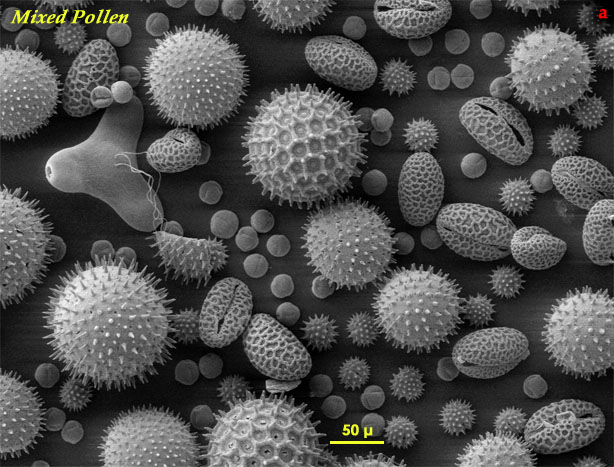
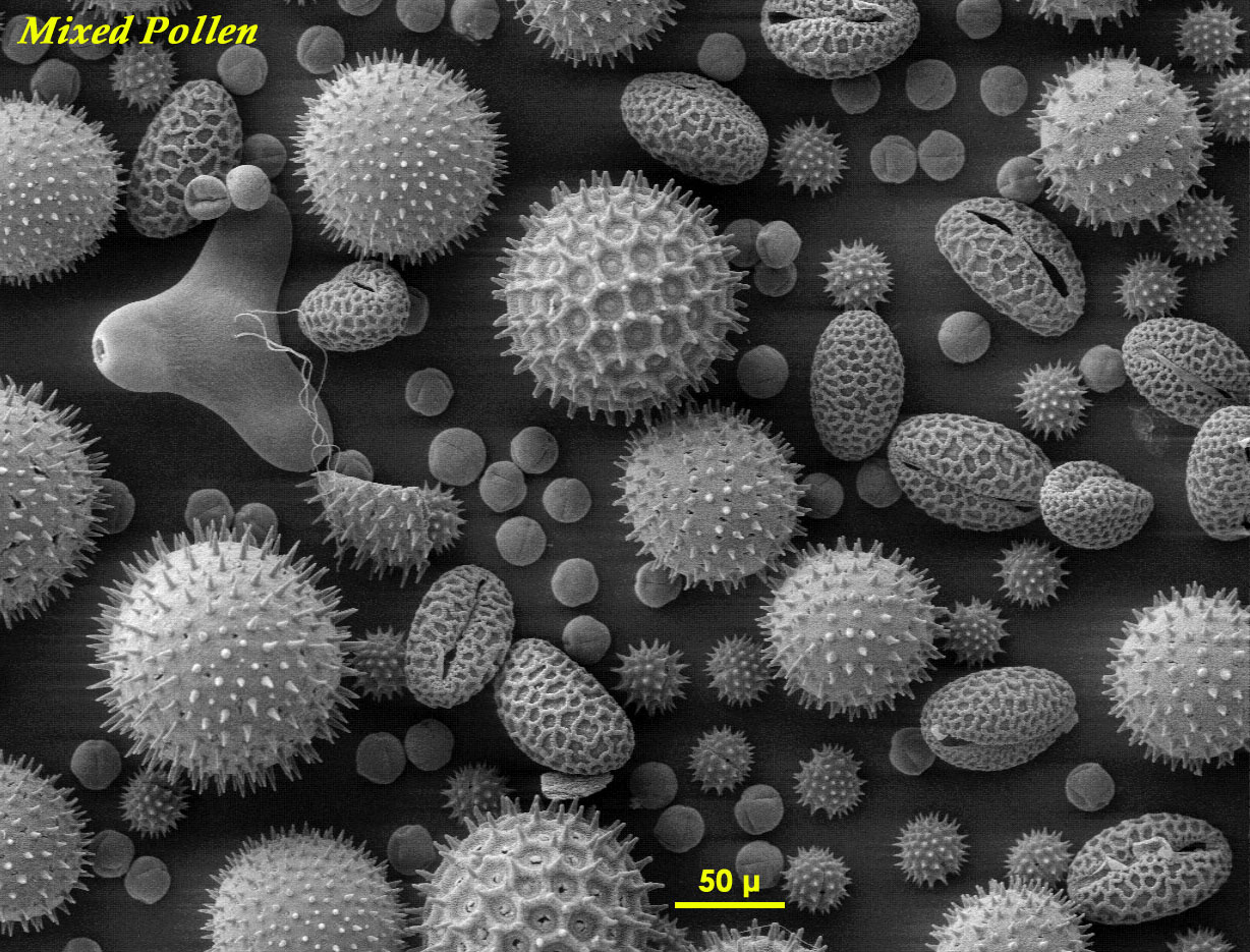
Although all pollen grains have the same function, they vary in shape, size, colour and in the architecture of the exine - the outer coating of the pollen grain. In recent years, the popular way to image pollen has been by scanning electron microscopy (SEM) which gives an excellent view of the exine: Fig.a is an example of pollen from a range of species imaged by this method. Though by no means comprehensive, it is a good example of how pollen can vary. Of course SEM gives no indication of colour, so another dimension of variation can be added to those already apparent in Fig.a. It should also be remembered that SEM involves intensive processing (high pressure, dehydration, high vacuum and metal coating) which may, sometimes, introduce structural distortions.
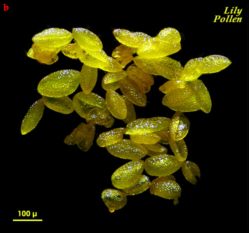
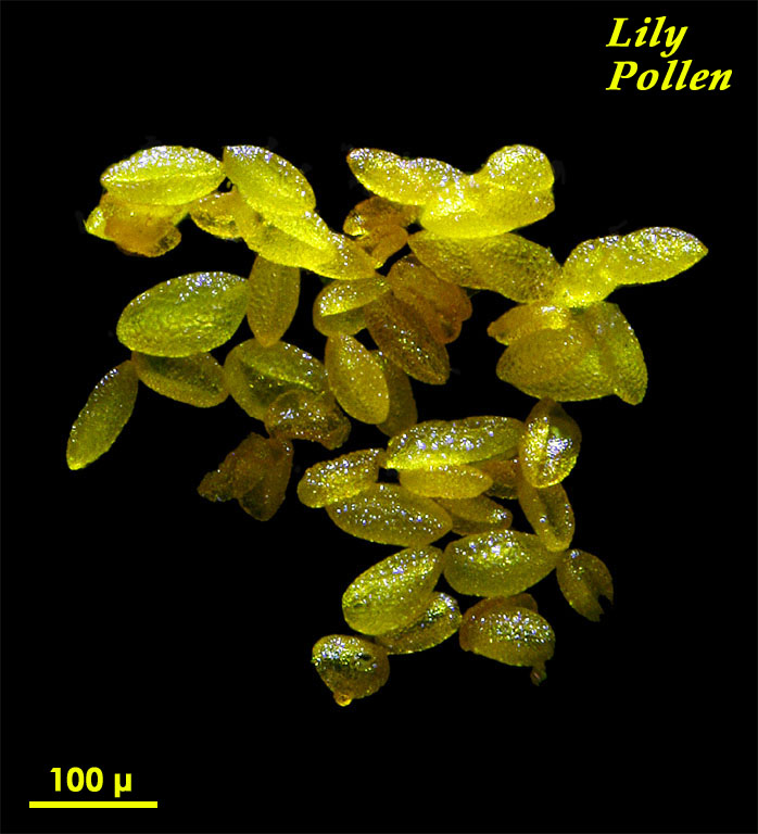
Fig.b shows live pollen grains of Lilium cv. imaged by dark ground incident light microscopy, which involves no processing whatsoever.
